 SINUSITIS
•Intracranial complications of acute bacterial rhinosinusitis include meningitis, brain abscess, subdural empyema and cavernous sinus thrombosis. Most common presenting feature of meningitis complicating sinusitis is seizure activity. •IDSA encourages use of antibiotics for acute bacterial sinusitis when strict diagnostic criteria are met. Amoxicillin/clavulinateremains first line therapy. NEONATAL VESICULAR RASH •Many causes of vesicular rashes in the neonate are benign, but serious bacterial and viral infection must be evaluated. •Neonatal scabies infestation presents with vesicular rash affecting the hands, feet, wrists and face of neonates. Treatment is with a single application of permethrin 5% cream, or 5-10% sulfur suspension in petroleum. PANCREATIC CANCER •Pancreatic cancer presents often in late stage due to vague symptoms of weakness, abdominal pain, diarrhea and jaundice. •Recurrent visits for similar chronic complaints carry high risk. Consider serious pathology as underlying source.
0 Comments
 Post-op Incisional Hernia
- Seen in over 10% of patients; up to 25% of patients with incision infection. - More common in midline incision, more common in upper abdomen vs lower abdomen. - Dr. Gibbs Pearl: If a patient presents to the ED with 30 days of their surgery (and is not there for an obviously unrelated complaint) contact the Surgeon to discuss the patient's presentation. - Surgery Pearl: evaluation for post-operative pain from lap chole can involve RUQ ultrasound to look for signs of abscess. Also consider biliary studies if concerned for biliary leak, biloma development. Negative CT Calcium score in ACS - Negative CT calcium score misses ACS very rarely. - Quick test that is non-invasive, has no contrast, does not require patient participation, does not rely on patient heart rate or ability to exercise. - If used in correct patient population, NPV is between 93-97% with a sensitivity of 99-100%. - Dr. Garvey Pearl: Recognize that your clinical gestalt trumps any protocol or clinical decision rule and do what you think is best for the patient. Missed Dialysis & Syncope
- PR prolongation, QRS widening - can be a full block - Sine wave
RUQ Abdominal Pain
**** Can have ectopic pregnancy with declining beta****
Missed Pubic Rami Fracture
**** Fully examine your patients and document well & make sure to reassess**** 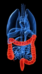 CORE CONCEPTS
•Incidence of CT scan ordering for IBD patients has more than doubled in the past 10 years. •Patients with Crohn’s disease are much more likely to have critical findings. •Patients with IBD are at increased risk of malignancy from radiation. •Take into account certain characteristics, ESR, & CRP when considering imaging. IBD Review Crohn's Disease - transmural inflammation, affects whole GI tract, skip lesions Ulcerative colitis - involves colon only Do We Image? --Yarur et al found that 93% of people with IBD in the ED with GI complaints had abnormal CT scan and in Crohn's disease 1/3 had clinically actionable findings and 12% of UC had clinically actionable findings. --Mo' CTs, Mo' Problems.. $$$$ - $1000 per CT scan Radiation - 1 in 1500 risk of death from malignancy from 50 mSv. --Keep in mind - IBD folks tend to be young with baseline increased risk of cancer from rapidly multiplying cells and immunsuppressive meds -- How to decide who to scan - Yarur et al - underweight, biologics use, previous IBD surgery, black race and HR > 90 were associated with increased risk for clinically actionable CT findings for Crohn's patients; salicylate use was protective. - proposed prediction models - PA+ model = ESR + (5xCRP); if less than 10, then no scan - 97% sensitivity Black Widow Spider Symptoms
LEADSymptoms
Cyclopeptite containing mushrooms
 CASE ONE: Epigastric pain after colonscopy Colonoscopy complications -Perforation - instrument vs air pressure -Hemorrhage -Visceral injury (spleen, diaphragm, volvulus) -Infection (bacteremia, retroperitoneal abscess, appendicitis) -Pneumatic injuries (Distention, pneumoperitoneum, pneumothorax) -Rate of perforation 1/1500... increases with biopsy/polypectomy Splenic laceration/rupture from colonscopy -Incidence 0.00005-0.017%. Likely under-reported. 14 Million colonscopies per yr in US -Mortality 5%. Danger lies in delayed diagnosis. Symptoms often attributed to air insufflation or serositis. -Female predominance, increased risk prev abdominal surgeries -Mechanism? Traction on splenocolic ligament, adhesions, direct trauma -Presentation: Abdominal pain, dizziness, Kehr's sign, worsening anemia (Kehr sign referred pain from diaphragm to shoulder) -Dx; Ultrasound, DPL, CT -Management: Symptomatic, serial Hb and abd exam, possible surgery CASE TWO: Sore throat Uvular Edema: Rarely reported in isolation Etiologies: Allergy/anaphylaxis, infection, angioedema, trauma Quincke's Disease: Inhalation injury, hereditary angioedema, cocaine/marijuana abuse, idiopathic/snoring in obesity. Presentation: Dysphagia, odynophagia, sore throat, foreign body sensation. Uncommonly resp distress, dyspnea, fever. -Some association with epiglottis -If infection suspect, cover strep species and consider lateral neck xray -If not infectious, steroids +/- H2 blockers -75% recurrence rate - either first 48 hours or remote CASE THREE: Groin pain Avascular necrosis of femur -Risk factors. Trauma, steroid use, hemoglobinopathy, dysbaric phenomena, autoimmune disease, storage disease, smoking, HLD, excess alcohol consumption -Often present with groin pain, throbbing, deep. Bilateral disease 50-80% -Can by identified by painful forced internal rotation at hip -Radiographic findings: Crescent sign (intact bone with deeper area of necrosis). On MRI "single density line" early, "double line sign" found in 80% cases, high intensity line surrounded by low intensity -Treatment: 85% collapse rate, surgical intervention is mainstay. Of those treated with conservative therapy, 76% proceeded to arthroplasty  TRAUMA PATIENTS
- CT if there is abdominal tenderness or a seatbelt sign - Seatbelt sign- Not just abrasions! They have bruising / ecchymoses. - With Seat Belt Sign, incidence of hollow viscus injuries (17%); splenic injuries (10%) - Things that are easy to miss on CT scan - Diaphragm injuries - think about in abdominal pain w/ dyspnea or chest pain - GI tract injuries - free fluid; bowel in discontinuity - Pancreatic injuries - difficult to tell difference between contusion and ductal injury - - Important because these need intervention and waiting will be detrimental; - ERCP or MRCP - definitive diagnosis ABDOMINAL PAIN IN THE ELDERLY - Have a low threshold for imaging elderly patients with abdominal pain - Abdominal pain in elderly - 60-70% admitted; 30% with surgical process; 10% with return ED visits; 5% mortality - Exam is not as reliable!! - CT: diagnostic in 85% of people who had emergent surgical process - Contrast? IV/ PO/ both/ CTA?- if concerned about vascular - get CTA > Contrast allergy - only true contraindication - airway compromise > CIN - Cr rising >0.5 mg/dL or 25% from baseline; > Most elderly people have risk factors Cr > 1.5-2.0 > Consider no contrast - if giving contrast - HYDRATE - Metformin > Manufacture warning - no metformin 48 hrs before or after IV contrast; > Increased risk - contraindications to metfomin, preexisting renal dysfunction BOWEL OBSTRUCTION - Diagnosis - Dilated loops of small bowel - diameter > 2.5 cm; - >50% difference in caliber before and after transition - Acute Obstruction Series X-rays - help if diagnostic; not so much if "normal" or "non-specific" - If an obstruction series is nondiagnostic and you suspect bowel obstruction, get a CT - PO contrast may add functional info, but often difficult to get into the patient... and not necessary. - PO contrast is almost never needed in CT scanning OTHERS - Diverticulitis - fat stranding; bowel wall thickening - CT with IV contrast best image - Appenditicits - senstivity 90% with no contrast; 100% with contrast - Mesenteric ischemia - CT angio is the best - Acute cholecystitis - cholesterol vs pigment stones can be seen on CT - Ultrasound is the first best test for suspected gallbladder pathology - Pancreatitis - Generally don't need imaging. - Consider imaging if changing clinical picture; not typical; no classic pancreatitis risk factors (to r/o malignancy)  CORE CONCEPTS
Special Circumstances:
1. Spearing injury - duodenal injuries, rectus injury with hernia, pancreatic injury - handlebar, ski pole, - duodenal hematoma- CT with oral contrast - pancreatic injury - get lipase (usually presents in delayed fashion) 2. Seatbelt sign - Ecchymosis / Bruises ... not just abrasions - get CT, give good return precautions or have them come back for check 3. Geriatric - VS misleading, abdominal exam insensitive, lactate > 2( sensitive to occult shock) - liberal role for CT - low threshold for admit PECARN Rules: get CT if, - Abdominal wall trauma, seatbelt sign - GCS< 14 - Tenderness - Chest wall trauma - Intoxication, - Hematuria - Elevated LFT - Painful distracting injury  Iron
- Metals in salt form cause VOMITING - 2+ ferrous sulfate in absorbable state → 3+ state for storage/transfer Chewable tablets - 10-18mg/tab - Hard to overdose on these - “minimally toxic” Iron carbonyl - elemental iron - low toxicity Iron filings in hand warmers → if ingested, could be toxic Prenatal vitamins - greatest morbidity - look like candy
Injuries
5 stages of toxicity 1. ingestion to 6h - vomiting!! - abd pain - diarrhea - melena/hematemesis - bowel wall necrosis/infarct 2. 6h - 24h - quiescent stage - symptoms appear to resolve - continued worsening acidosis - if ingestion was small → course usually stops here - if ingestion was large → this stage is sometimes skipped and go onto more badness 3. 12h - 48h - crash - CV, Liver, GI, ARDS, CNS lethergy/coma, Acidosis 4. 2d - 3d - independent of severity of stage 3 - fulminant hepatic failure - >1000 iron level 5. weeks later - mucosal injuries/strictures Iron Levels
Workup - electrolytes -- AGMA - coags if bleeding - LFTs if sick - APAP level for intentional ingestion -- think about synergy - x-rays -- abd → see pills sometimes Management
use: hx of sxs, pos xrays, super high iron level 100mg binds 10mg iron IV admin -- 15mg/kg/hr for rate but may not get in enough Side Effects: hypotension, tachycardia, diuresis visual/ototoxicity, abd pain, fever, diarrhea increased risk for yersenia enterocolitica sepsis stop when acidosis resolves Core Concepts: elemental dose is what’s toxic no charcoal for tx look for anion gap metabolic acidosis check an xray for pills, but if it’s negative doesn’t mean pt isn’t sick there’s a quiescent phase of toxicity deferoxamine is an option for iron chelation pay attention to units used to quantify iron --usually in dL  DDX of projectile vomiting
Imaging US - muscle length >17mm positive, width >3mm positive - tips: need true sag, need relaxed muscle UGI - If US not available - string sign |
Archives
August 2018
Categories
All
|
 RSS Feed
